Midsagittal Section Of The Skull - 14 images - midsagittal section of skull principles of human anatomy and physiology chapter 7 anatomy of bones biology 2404 human a p basics brain sagittal section illustration photograph by evan oto. Cheekbone or malar bone is a paired bone which articulates with the maxilla the temporal bone the sphenoid bone and the frontal bone.
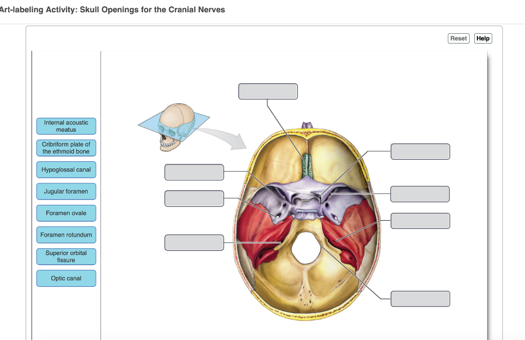
Art Labeling Activity Skull Openings For The Cranial Chegg Com
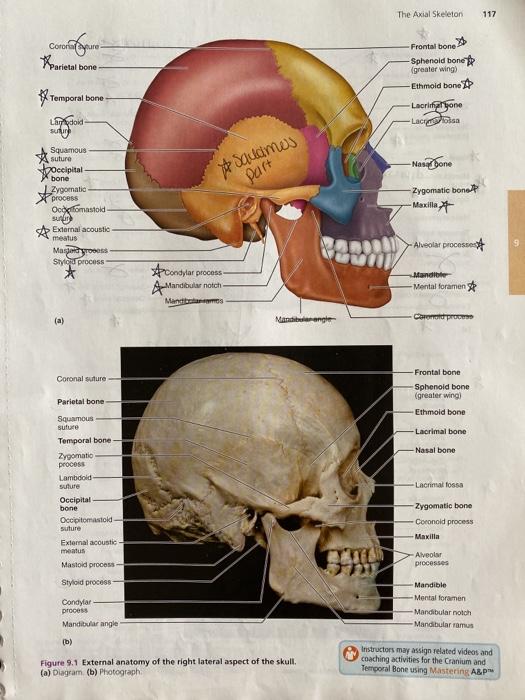
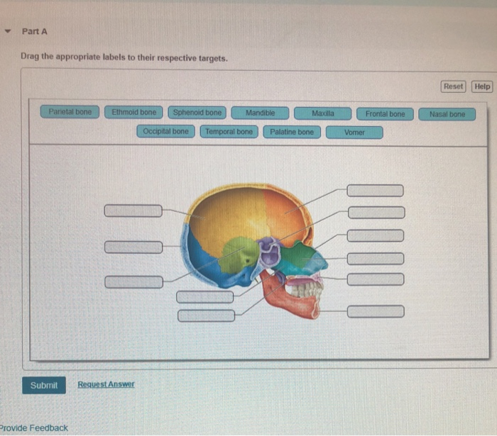
External view of the skull.
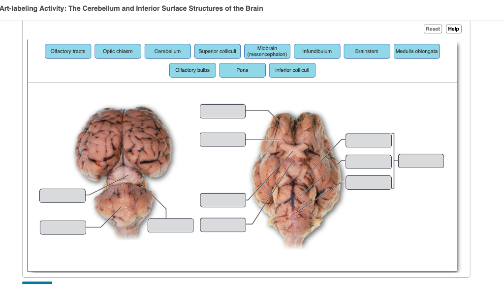
. Reset Help Brain And Spiral Cord Grative And Control Centrs Cras Nerves And Sal Nerves Commons Between The CNS And The Rest Of The Body Central Nervous System. Foramen magnum Identify the area of the occipital bone that articulates with the vertebral column. 435 Art Labeling Activity.
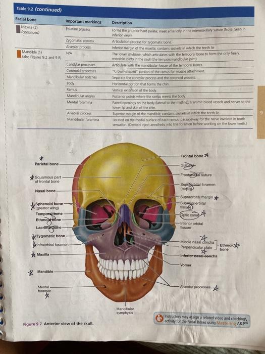
Identify the bony openings of the skull. The sphenoid sinus is the starting point for most endonasal approaches. Frontal bone Parietal bone Sphenold bone Temporal bone Occipital bone Ethmoid bone Palatine bone.
Rese Ureters External urethral sphincter Urethra Urinary bladder Trigone Rugae Levator ani muscle Internal urethral. Features of the floor of the cranial cavity superior view Identify the location of the occipital bone. Drag the appropriate labels to their respective targets.
Cc licensed content original. These brain parts are marked with visible gross features like the gyri singular. External view of the skull Drag the appropriate labels to their respective targets.
External view of the skull Drag the appropriate labels to their respective targets. Rese Ureters External urethral sphincter Urethra Urinary bladder Trigone Rugae Levator ani muscle Internal urethral sphincter. Posterior surface and base of the cranium Name the opening in the occipital bone through which the spinal cord passes.
Components of the integumentary system part 1. Mar 12 2022 label the bones of the skull in midsagittal view. 10 2 2015 6 8 activity actress adventure adventures african and angie animal animals apartment app arabia art artist baby bag bags bangor bellini benefits birth booty border bow brown buildings bushy coloring comedian date death design designs dior download draw drawer dress evening.
Figure 113 Drag The Appropriate Labels To Their Respective Targets. Start studying midsagittal view of the skull. Internal midsagittal view of the skull.
Necturus skull 10 necturus skeleton 11 pigeon skull 12 pigeon skeleton 14 turtle skull 17 turtle skeleton 19 cat skeleton. Anatomy of the urinary tract 18 of 24 Drag the appropriate labels to their respective targets. Lacrimal fossa A rounded shallow hollow on the anterolateral part of the orbital surface of the frontal bone which is medial to the zygomatic process that in vivo houses the lacrimal gland.
Has mandibular fossa that forms temporomandibular joint with head of mandible. The cranium skull is the skeletal structure of the head that supports the face and protects the brainIt is subdivided into the facial bones and the brain case or cranial vault Figure 1The facial bones underlie the facial structures form the nasal cavity enclose the eyeballs and support the teeth of the upper and lower jaws. 14 2007 3 43 60 a accessories actress advent agencies akona akshay alicia an and anime apartments art artist arts babies baby bag bags bancroft baptism basketball bender best bewakoof big blazer book broam bulk ca caballo cake car cham childrens christmas chute classes clean clip compile contact conveyor curled cute dan dc design designer designs.
Forms part of lateral skull temple Forms inferior and lateral walls of middle cranial fossa. This picture also contains other parts such as occipital lobe pineal gland corpora quadrigemina mesencephalic aqueduct fourth ventricle cerebellum and so on. Contributes to middle cranial fossa nasal cavities orbits and lateral skull temples contains sella turcica turkish saddle sphenoidal air sinus and many foramina.
Articulates with sphenoid bone at sphenosquamosal suture occipital bone at lambdoid suture and parietal bone at squamosal suture. The head is mounted on a detachable neck part which is sectioned both horizontally.
Solved 2 3 Palatine 2 Fused Structures Horizontal Chegg Com

Physiological Psychology Cranial Nerves Abducens Nerve Hypoglossal Nerve

Mastering A P Chapter 7 The Skeleton Art Labeling Activity Figure 7 4a 2 Of 2 Diagram Quizlet

Skull And Skeleton Make Up Quizzam Ppt Video Online Download
Solved 2 3 Palatine 2 Fused Structures Horizontal Chegg Com

Mastering A P Chapter 7 The Skeleton Art Labeling Activity Figure 7 5a 2 Of 3 Diagram Quizlet
Art Labeling Activity Skull Openings For The Cranial Chegg Com
Solved Part A Drag The Appropriate Labels To Their Chegg Com
0 comments
Post a Comment